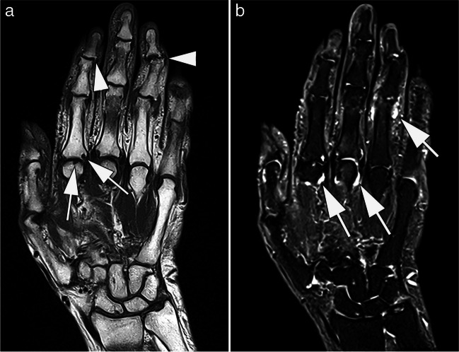
Fig. 3.
MRI at 0.55 T of the hand of a 77-year old patient with suspected late-onset rheumatoid arthritis. Coronal T1-weighted (A) image shows erosions at the 2nd metacarpophalangeal joint (arrows). In addition, osteophytes are depicted at the 2nd and 4th distal interphalangeal joints (arrowheads). B Corresponding STIR image shows synovitis at the metacarpophalangeal joints 2 and 3, and at the proximal interphalangeal joint 4 (arrows). TA: 03:20 min (A), 05:11 min (B)

