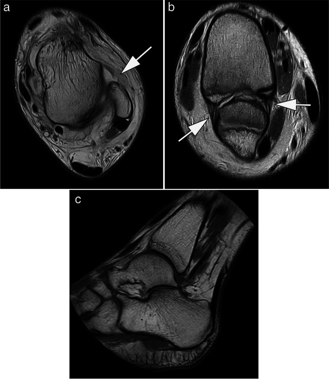
Fig. 6.
MRI at 0.55 T of the ankle of a 14-year-old patient after ankle sprain. A Axial proton density-weighted image shows a complete rupture of the anterior talofibular ligament (arrow). B Parasagittal proton density-weighted image demonstrates that the anterior and posterior tibiofibular ligaments are intact (arrows). C Small-bore dedicated 1.0-T extremity MRI allows for assessment of the ankle joint, but depending on size of the patient usually plantar flexion is applied as shown in this T1-weighted spin echo image. Note circular field of view of magnet system. TA: 03:07 min (A), 05:26 min (B), 04:21 min (C)

