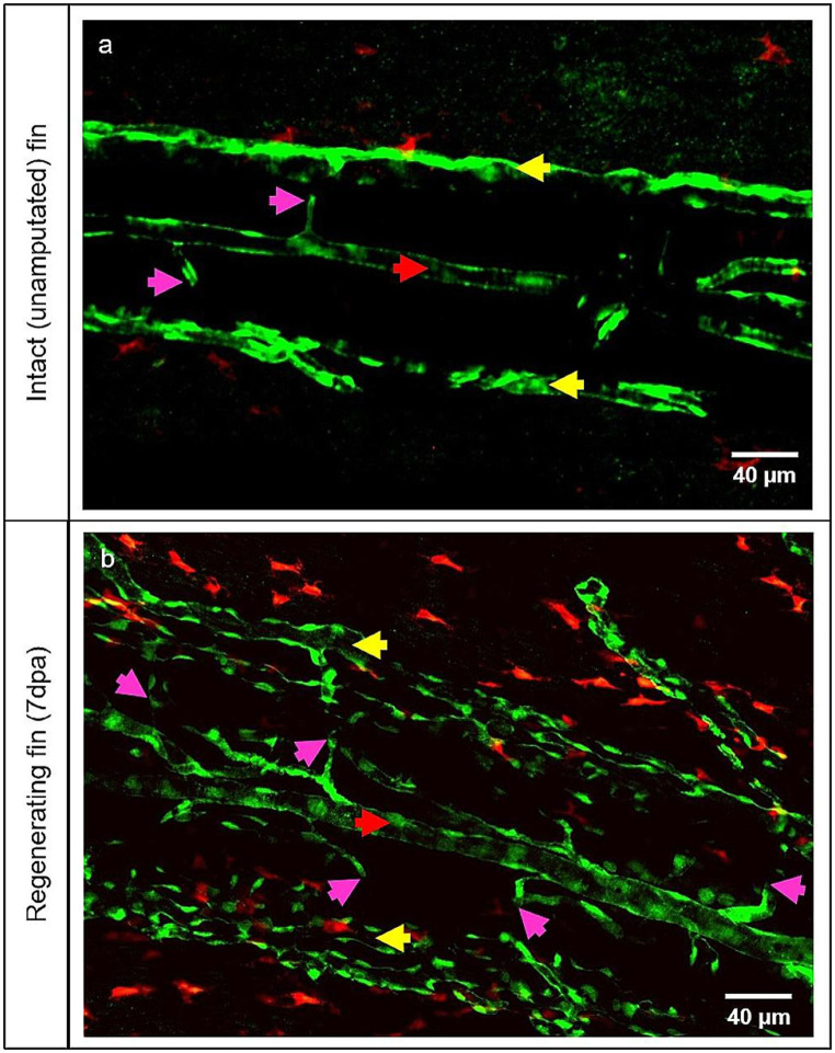
Fig. 3.
MΦ augmentation during the caudal fin regeneration. In vivo appearance of MΦ by the intact (unamputated) fin (a) and regenerating fin at 7dpa (b). Blood vessels (ECs) appear in green and MΦ in red (transgenic line). (a) displays hierarchically simple-organized blood vessels containing an artery (red arrow), veins (yellow arrows) and connecting capillaries (purple arrows), and only a few tissue MΦ. (b) displays a dense, complex blood vessel network with multiple MΦ surrounding the blood vessels and scattered in the tissue in between. Right side – zebrafish tail; images acquired by confocal microscopy

