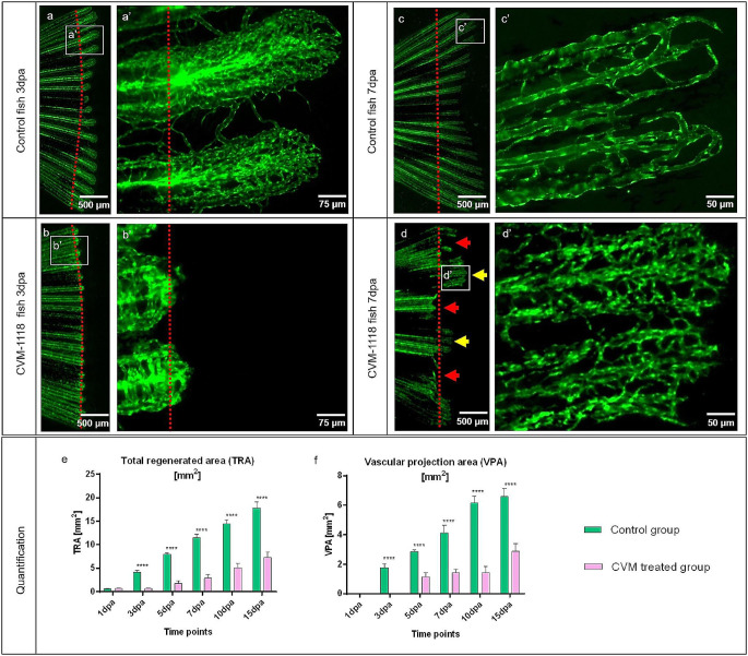
Fig. 5.
Inhibition of vascular mimicry by CVM-1118 impairs caudal fin regeneration and blood vessel formation. Vascular changes during normal fin regeneration (a, c) versus regeneration in the presence of CVM-1118-treated group (b, d) at 3dpa and 7dpa (green, transgenic zebrafish line; red dash line – amputation plane). Blood vessels of the controls (a, a’ and c, c’) appear well organized and represent the classical vascular regeneration pattern. b and b’ display shorter regeneration area with respectively modest vascularization in treated animals; hierarchical vessels and capillaries are less pronounced. At 7dpa, the regenerating tissue is severely impaired; smaller vascular plexus and regeneration processes, including blood vessel formation, is completely absent in half of the fin area (d, red arrows) in comparison to the control. In the partially regenerated regions (d, yellow arrows), tissue and the vascular plexus appear shorter with a dense and poorly organized capillary meshwork (d’). Images are acquired by fluorescent reflected light microscopy. Quantification of the tissue regeneration and vascularization after inhibition was assessed by two variables: Total regenerated area (TRA = regenerated fin in mm2; (e) and vascular projection area (VPA = vessels within regenerated part in mm2; f) during the period of 15 days in control group (green) versus CVM-1118 treated group (purpura); n = 5. In the treated group, TRA and VPA are significantly smaller (about 70%); fin shape and size never returned to the amputated stages indicated by control

