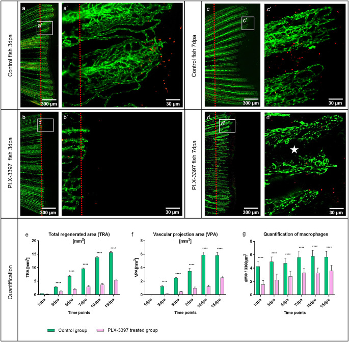
Fig. 7.
Macrophage inhibitor PLX-3397 impairs caudal fin regeneration, blood vessel formation and macrophage appearance. Vascular alteration during normal fin regeneration (a, c) versus PLX-3397-treated group (b, d) at 3dpa and 7dpa is documented (green – ECs, red – MΦs; transgenic zebrafish line; red dash line – amputation plane). In the control animals, classical tissue regeneration pattern, blood vessel morphology and MΦs are documented (a, c). Regenerative area and vascular plexus is severely impaired and underdeveloped in the PLX-3397-treated group (b, b’) at 3dpa; only a few capillaries are observed and less MΦs detectible. In the treated group, at 7dpa, the regenerating region and the vascular plexus are smaller, modest and capillaries are less pronounced (d, d’). Intraray region is not properly vascularized (d’, white asterisk) and less MΦs are observed in comparison to the control group. Images are acquired by fluorescent reflected light and confocal microscopy. Quantification of the regeneration and vascularization after the MΦ elimination has been performed by two variables: total regenerated area (TRA = regenerated fin in mm2; (e), vascular projection area (VPA = vessels growth within regenerated fin in mm2 (f)) during the period of 15 days in control group (green) versus PLX-3397-treated group (purpura). n = 5. (h) Quantification of MΦ amount between control and PLX-3397-treated animals; n = 100. In the treated group, TRA and VPA are significantly smaller (about 75%), never reaching the parameters of the controls (e, f); Amount of MΦ is significantly reduced (about 50%) in the PLX-3397-tretaed animals during the entire course of experiment in comparison to control animals (g)

