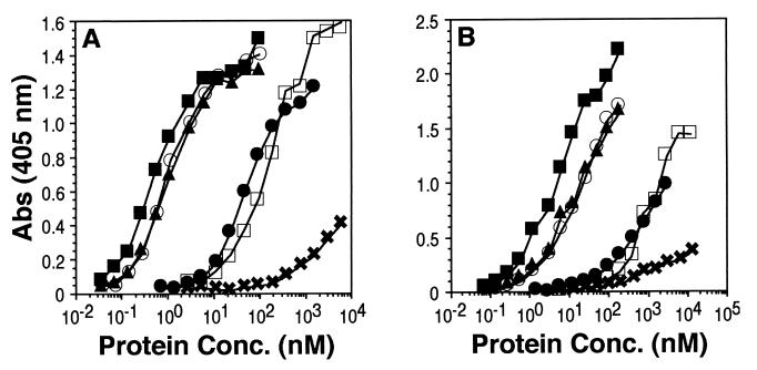
FIG. 3.
Analysis of gD truncation mutants for receptor binding by ELISA. The wells of an ELISA plate were coated with an excess of HveA(200t) or HveC(346t) and incubated with increasing concentrations (shown on the x axis) of gD truncation mutants. Bound gD was detected by incubating sample wells with a rabbit antiserum raised against gD (R7), followed by peroxidase-conjugated goat anti-rabbit antibody and then peroxidase substrate. (A) Binding to HveA(200t). (B) Binding to HveC(346t). Symbols: □, gD-1(306t); ■, gD-1(285t); ▴, gD-1(260t); ○, gD-1(250t); ●, gD-1(240t); ✖, gD-1(234t).

