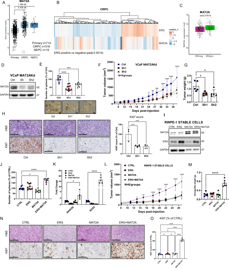
Fig. 1. MAT2A is elevated in aggressive prostate tumors and its ablation reverts transformation in vitro and in vivo.
A Gene expression level of MAT2A in prostate cancer patients indicated subgroups (Primary, n = 714; CRPC, n = 316; NEPC, n = 19). p-values are indicated. B Heatmap of ERG and MAT2A expression values in the cohort of CRPC patients described in A. (Padj value 0.0014). C Gene expression level of MAT2A in CRPC described in A and divided in ERG positive (n = 113) and negative (n = 203). Adjusted p-value 0.0014. D Immunoblot of VCaP control (Ctrl) and MAT2Akd VCaP cells (Sh1, Sh2) with indicated Ab (n = 3 independent experiments). E Sphere formation assay (SFA) in indicated cell lines (n = 3 independent experiments with n = 4 (for sh1) and n = 3 (for ctrl and sh2) biological replicates. Bottom, representative images of tumor spheres. Scale bars, 100 µm. F Growth curve of VCaP xenograft from VCaP control (Ctrl) and VCaP with stable MAT2A knockdown (Sh1 and Sh2) were engrafted and growth was monitored by caliper every 2 days (n = 5 mice /group). G Tumor weight of indicated tumor xenografts (n = 5 mice/group). H Representative sections from the indicated tumor xenografts. Scale bars, 200 µm. Right, Immunoscore of Ki67 by Aperio tool (n = 3 mice Ctrl, n = 5 mice Sh1, n = 5 mice Sh2). I Immunoblot of RWPE-1 cells with stable expression of ERG, MAT2A and ERG + MAT2A with indicated Ab (n = 3 independent experiments). J SFA in indicated cell lines (n = 3 independent experiments with 3 biological replicates). K Fold change of mRNA levels of NANOG and SOX2 in RWPE-1 cells with stable expression of ERG, MAT2A and ERG + MAT2A. (n = 5-6 biological replicates). L Growth curve of RWPE-1 xenografts. Indicated cell lines were engrafted and growth was monitored by caliper every 2 days (n = 6 mice/group). M Tumor weight of indicated tumor xenografts (n = 6). N Representative sections from the indicated tumor xenografts. Scale bars are 200 µm. O Immunoscore of Ki67 using the Aperio tool (n = 6 mice/group). Molecular weights are indicated in kilodaltons (kDa). All error bars, mean ± SD. For box-and-whisker plots in A and C, the line inside the box shows the median value. The bounds of the box represent the 25th–75th percentiles, with whiskers at minimum and maximum values. One-way-ANOVA was used to test significant differences between groups in all panels, except for (K) where 2-way-ANOVA was used. Data presented in F and L are independent replicates derived from individual mice. *p < 0.05, **p < 0.01, ***p < 0.001 ****p < 0.0001.

