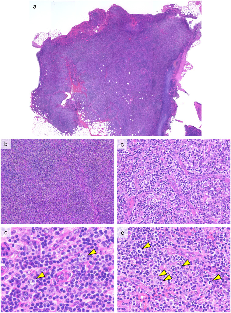
Fig. 2.
Histopathological features of ALPIBP (Case 1: H&E staining)
a, b: The follicular structure is obscured and the interfollicular area is expanded.
c: Vascular proliferation with plump endothelial cells is seen in the expanded interfollicular area.
d: Immunoblasts with large nuclei and distinct nucleoli (arrow heads) are observed with a background of small lymphocytes and plasma cells.
e: Mitotic figures (arrow heads) are easily observed.

