Figure 1.
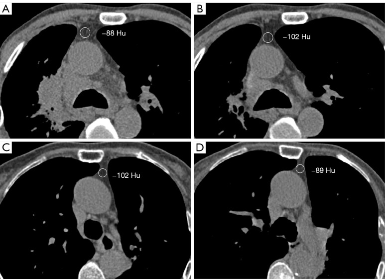
Measurement of thymic density in the anterior mediastinum: (A,B) the first and last chest CT images of the same patient, respectively, who showed an annual decrease in thymic density; (C,D) the first and last chest CT images, respectively, of another patient, who presented with an increase in annualized thymic density. HU, Hounsfield unit; CT, computed tomography.

