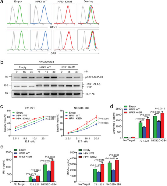
Figure 4.

HPK1 overexpression and its kinase activity mediate human NK cell dysfunction. a) Establishment of NKL cell line expressing HPK wild‐type (WT) and its kinase‐dead mutant (K46M) by retroviral transduction. NKL cells transduced with the retrovirus containing sequences for WT or K46M form of HPK1 were sorted for a matched level of GFP expression. The level of HPK1 protein as reflected by GFP expression in the transduced cells was measured by flow cytometry. Representative flow cytometric analysis of HPK1 expression (top) and GFP expression (bottom) in NKL cells transduced with empty vector (green), HPK1 WT (blue), or HPK1 K46M (red). The retroviral construct without an HPK1 sequence (empty) was used as a control. b) NKL cells expressing HPK1 WT or HPK1 K46M were stimulated through a combination of NKG2D and 2B4 for the indicated times. Cell lysates were immunoblotted for phospho‐SLP‐76 at serine 376 (pS376), HPK1, or total SLP‐76. c) Lysis of 721.221 (left) or P815 cells engaging NKG2D and 2B4 (right) by NKL cells expressing HPK1 WT or HPK1 K46M at the indicated effector to target (E:T) cell ratio, as determined by europium assay (triplicate samples per group). d) NKL cells expressing HPK1 WT or HPK1 K46M were stimulated with 721.221 cells or a combination of NKG2D and 2B4 for 2 h. The secretion of granzyme B in the supernatant was measured by ELISA (triplicate samples per group). e) NKL cells expressing HPK1 WT or HPK1 K46M were stimulated with 721.221 cells or a combination of NKG2D and 2B4. After 8 h of incubation, IFN‐γ (left) and MIP‐1α (right) released in the supernatant were measured by ELISA (triplicate samples per group). Data represent the mean ± SD (c, d, and e) and were analyzed using two‐way ANOVA with Dunnett's multiple comparison test (c) and two‐tailed unpaired t‐test (d and e); actual p‐values are indicated. All data are representative of at least 3 independent experiments.
