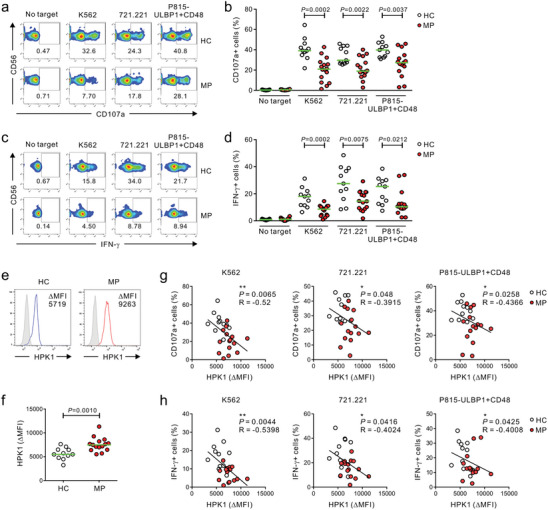
Figure 6.

Dysregulated HPK1 upregulation correlates with peripheral NK cell dysfunction in melanoma patients. PBMCs from the healthy control (HC) group (n = 11) and melanoma patients (MP) group (n = 16) were incubated with conventional K562 or 721.221 target cells, or P815‐ULBP1+CD48 cells that activate NK cells via NKG2D and 2B4. a,b) Cytotoxic degranulation of NK cells showing the representative result (a) and graph (b), as measured by the percent increase of surface expression of CD107a. c,d) Cytokine production by NK cells showing the representative result (c) and graph (d), as measured by percent increase of intracellular expression of IFN‐γ. e,f) Flow cytometric analysis of HPK1 expression in NK cells from HC group (n = 11) and MP group (n = 15) relative to isotype control (ΔMFI) showing the representative result (e) and graph (f). g,h) The expression of HPK1 (ΔMFI) in NK cells correlates inversely with the percentages of CD107a‐ (g) or IFN‐γ‐positive NK cells (h) after stimulation with all 3 target cells. Data were pooled from 5 independent experiments. Horizontal bars (green) indicate the medians (b, d, and f); each dot represents an individual donor. Data were analyzed using the Mann‐Whitney U‐test (b, d, and f) and Spearman correlation test (g, h); actual P‐values are indicated.
