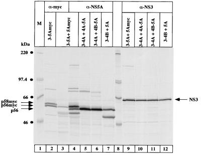
FIG. 4.
Analysis of NS5A hyperphosphorylation with NS5A and NS3 expressed in trans. Cells were cotransfected with the following plasmids: lane 2, pcD3-5Amyc alone; lanes 3, 4, and 9, pcD3-5A plus pcD5Amyc; lanes 5 and 10, pcD4A-5A plus pcD3-4A; lanes 6 and 11, pcD4B-5A plus pcD3-4A; lanes 7 and 12, pcD3-4B plus pcD5A. Proteins were immunoprecipitated with Myc-specific monoclonal antibodies (α-myc) NS5A antiserum (α-NS5A), or with NS3 antiserum (α-NS3) and loaded onto an SDS–7.5% polyacrylamide gel. NS5A p56, p58, and NS3 are indicated by arrows on the right; p56-Myc and p58-Myc are indicated by arrows on the left. M, molecular weight marker proteins.

