
Fig. 2.
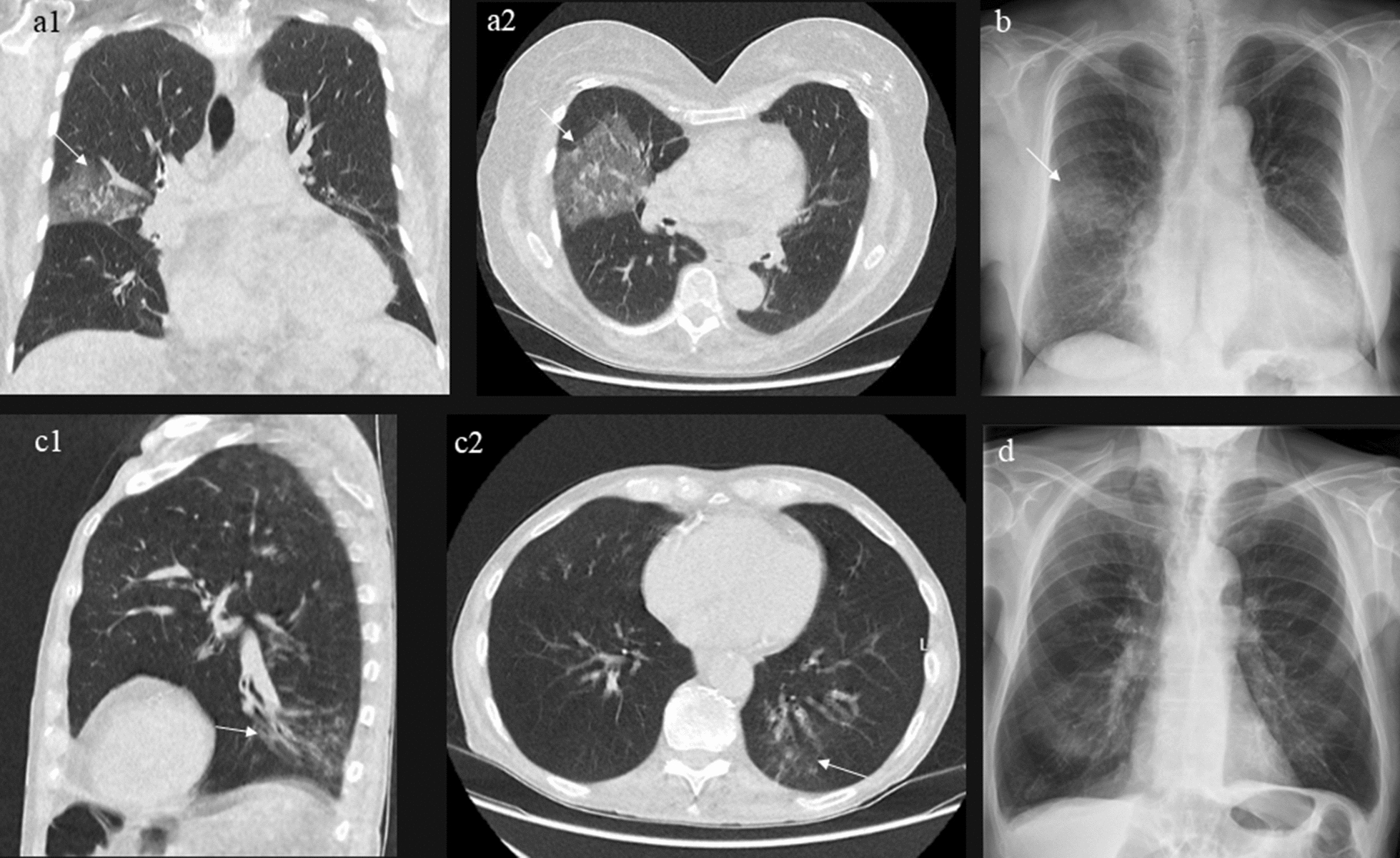
Examples of images from the study population (a) Ultralow-dose computed tomography (ULD-CT) of the chest from a patient with an opacity consistent with community-acquired pneumonia (CAP) in the right lobe, which is also seen in the corresponding chest radiograph (b). (c) ULD-CT of the chest from a patient with a left lower lobe opacity consistent with CAP. This opacity was not initially identified on the corresponding chest radiograph by the reporting radiologist (d). After reviewing the ULD-CT and comparing it with the chest radiograph, a small pneumonia could be suspected upon a second examination of the images (c, d)