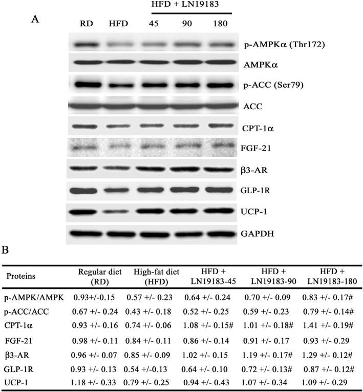
Fig. 2.
(A) Representative immunoblot images showing protein expressions of p-AMPK and AMPK, p-ACC and ACC, CPT-1α, β3-AR, GLP-1R, and FGF-21 in liver and UCP-1 in inguinal fat tissue lysates of the experimental groups of rats as indicated. (B) The table presents group-wise mean ± SD (n = 8) of the normalized protein expressions obtained from the densitometry analysis of the immunoblot protein bands. p-AMPK and p-ACC protein expressions were normalized with the respective un-phosphorylated proteins, and CPT-1α, β3-AR, FGF-21, GLP-1R, and UCP-1 were normalized with GAPDH. RD: regular diet, HFD: high-fat diet; # indicates significance (P < 0.05) in HFD vs. LN19183-supplemented with 45, 90, or 180 mg/kg/day groups comparison analyses using one-way ANOVA with suitable post-hoc test.

