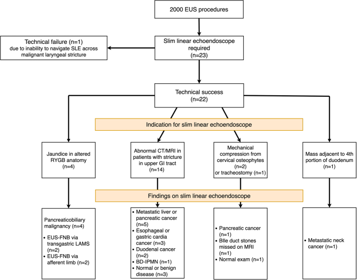
FIGURE 1.
Flow diagram of patients who failed EUS using standard echoendoscopes and required examination using SLE. CT indicates computed tomography; EUS, endoscopic ultrasound; FNB, fine needle biopsy; GI, gastrointestinal; IPMN, intraductal papillary mucinous neoplasm; LAMS, lumen-apposing metal stents; MRI, magnetic resonance imaging; SLE, slim linear echoendoscopes.

