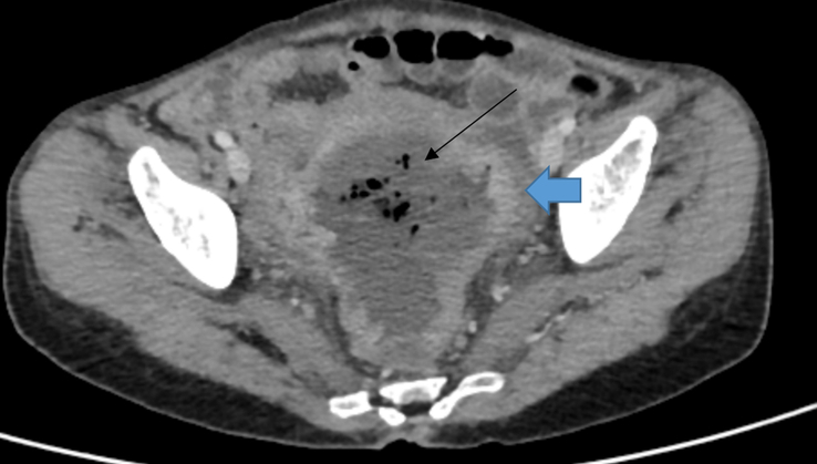
Figure 1.

Axial section of contrast-enhanced computed tomography (CECT) of abdomen and pelvis showing heterogeneously enhancing asymmetrical circumferential thickening of the proximal rectum and rectosigmoid junction (blue bold arrow) and dilated rectum with heterogeneous collection (black thin arrow).
