Figure 9.

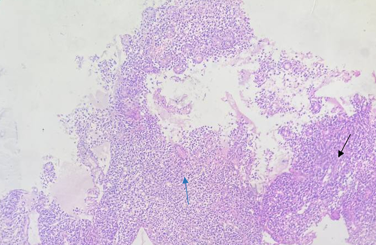
Photograph taken during histopathological examination of rectal biopsy, showing poorly. differentiated adenocarcinoma (Black thin arrow showing growth/mass and blue thin arrow showing mitosis).

Photograph taken during histopathological examination of rectal biopsy, showing poorly. differentiated adenocarcinoma (Black thin arrow showing growth/mass and blue thin arrow showing mitosis).