. 2024 Jul 19;25(8):726–741. doi: 10.3348/kjr.2024.0105

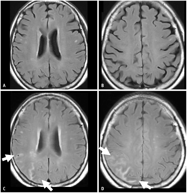
Fig. 4. A 69-year-old female patient presented with mild cognitive impairment and was treated with gantenerumab therapy. A, B: Her baseline MRI shows a mild degree of white matter hyperintensity. C, D: Seven months after the initiation of gantenerumab therapy, the patient had an episode of syncope, and the MRI showed multifocal sulcal FLAIR high-signal intensity in the right parietal lobe, suggesting ARIA-E (arrows). The case had two regions (arrows) with an extent measuring between 5 cm and 10 cm, classified as moderate. As a symptomatic and moderate case, the gantenerumab therapy was suspended. ARIA-E was resolved after suspending treatment for 1 month.

Fig. 4