. 2024 Jul 19;25(8):726–741. doi: 10.3348/kjr.2024.0105

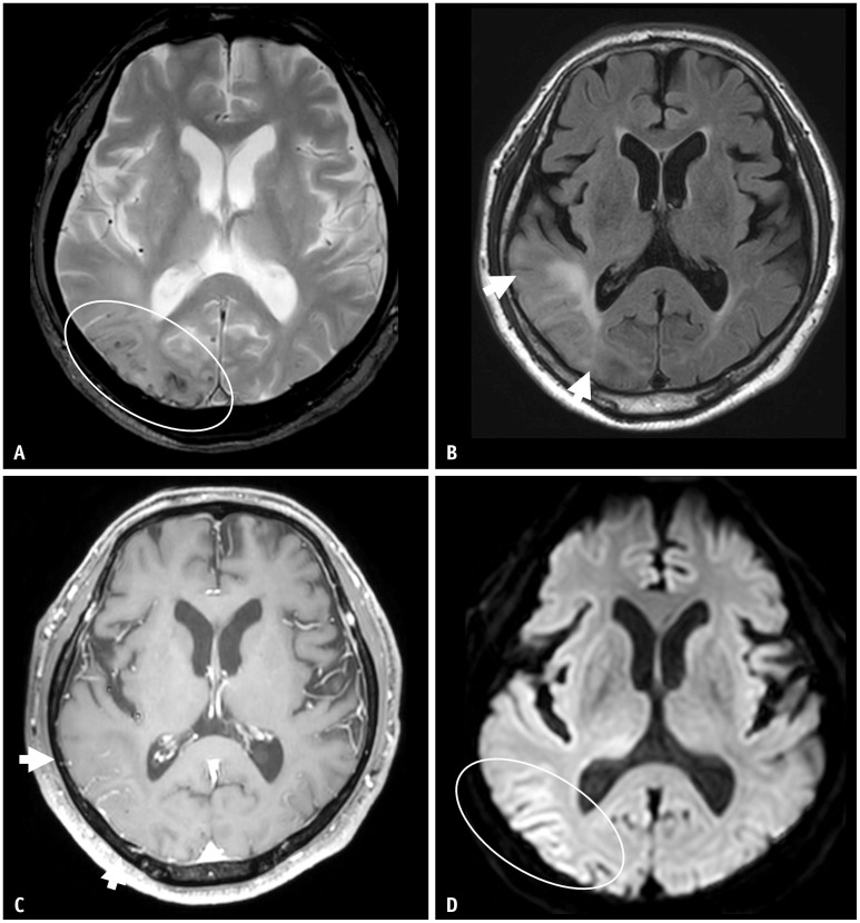
Fig. 7. A 70-year-old male patient presented with confusion. A: On T2* gradient echo sequence, there were multiple lobar distributed cortical and subcortical microbleeds in the right temporooccipital lobes. B: On FLAIR, there was confluent parenchymal high signal intensity (arrows). C: Postcontrast axial T1 images demonstrated marked gyral enhancement in the same area (arrows), suggestive of prominent leptomeningeal vessels. D: On the diffusion-weighted image, there was subtle high signal intensity along the cortex. These images are very similar to amyloid-related imaging abnormalities; however, the patient had not undergone anti-amyloid-beta immunotherapy. The patient was diagnosed with cerebral amyloid angiopathy-related inflammation.

Fig. 7