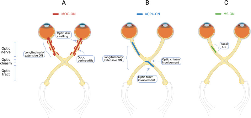
Fig. 1. Localisation of visual pathway involvement in MOG-ON, AQP4-ON and MS-ON.
A MOG-ON (red): Typical features include bilateral longitudinally extensive ON, with optic disc swelling, involvement of the retrobulbar segment of the optic nerve, and optic nerve sheath involvement or optic perineuritis. B AQP4-ON (blue): Longitudinally extensive ON, with optic chiasm +/− optic tract involvement. C MS-ON (green): focal ON. AQP4 aquaporin 4, MOG myelin oligodendrocyte glycoprotein, MS multiple sclerosis, ON optic neuritis.

