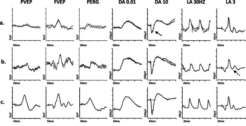
Fig. 3. Electrophysiological findings in SSBP1- and ALPK1- related pathologies.
Examples of pattern reversal VEPs (PVEP), flash VEPs (FVEP), pattern ERGs (PERG) and full-field ERGs are shown from one eye of a patient with (a) SSBP1-related pathology; b ALPK1-related pathology (ROSAH syndrome); and (c) from a representative normal subject for comparison. Recordings showed a high degree of inter-ocular symmetry and are shown for one eye only. All patient traces are superimposed to demonstrate reproducibility. The DA 0.01, DA 10 and LA3 ERGs include a 20 ms pre-stimulus delay. The patient with SSBP1-related disease (a) shows a subnormal PVEP, a borderline FVEP and reduction in the PERG N95:P50 ratio, with additional P50 peak time shortening, in keeping with macular retinal ganglion cell dysfunction; the DA ERGs are subnormal, with reduction (solid arrow) in the DA10 a-wave, which localises dysfunction at the level of the photoreceptors; LA ERGs are borderline normal. The patient with ALPK1-related pathology (b) shows a subnormal and delayed PVEP, a normal FVEP and reduction in the PERG N95:P50 ratio, in keeping with macular retinal ganglion cell dysfunction; the DA ERGs are borderline normal; LA ERGs are subnormal, indicative of generalised cone system dysfunction, with additional PhNR attenuation (arrow with broken lines), suggestive of global retinal ganglion cell loss.

