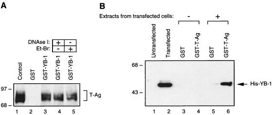
FIG. 2.
In vitro interaction between YB-1 and T-antigen. (A) Bacterially expressed GST (lane 2) or GST–YB-1 (lane 3) was immobilized on GST-Sepharose beads and incubated with whole-cell extract prepared from hamster glial cells constitutively expressing T-antigen (HJC-15b). Additionally, whole-cell extracts from HJC-15b cells, treated with DNase I (0.2 U/μg of protein) or ethidium bromide (100 ng/ml) (Et-Br), were also incubated with GST–YB-1 (lanes 4 and 5, respectively). HJC-15b whole-cell extract was loaded as a migration control (lane 1). Nonbinding proteins were removed from the column by extensive washing, and proteins interacting with GST or GST–YB-1 were resolved by SDS–10% PAGE and analyzed by Western blotting with an anti-T-antigen antibody (Ab-2 416). The bracket indicates T-antigen (T-Ag). (B) Whole-cell extracts prepared from untransfected HJC-15b cells (lanes 1, 3, and 4) and from HJC-15b cells transfected with a histidine-tagged YB-1 expression plasmid (pEBV-YB-1) (lanes 2, 5, and 6) were incubated with either GST alone (lanes 3 and 5) or GST–T-antigen (GST-T-Ag) (lanes 4 and 6) as indicated. After being washed, proteins interacting with GST or GST–T-antigen were resolved by SDS–10% PAGE and analyzed by Western blot analysis with anti-T7 antibody for detection of His-tagged YB-1. Whole-cell extracts from HJC-15b cells either untransfected (lane 1) or transfected with pEBV-His-YB-1 expression plasmid (lane 2) were loaded as negative and positive migration controls, respectively. The arrow indicates histidine-tagged YB-1. The positions of molecular mass markers (in kilodaltons) are shown on the left of each panel.

