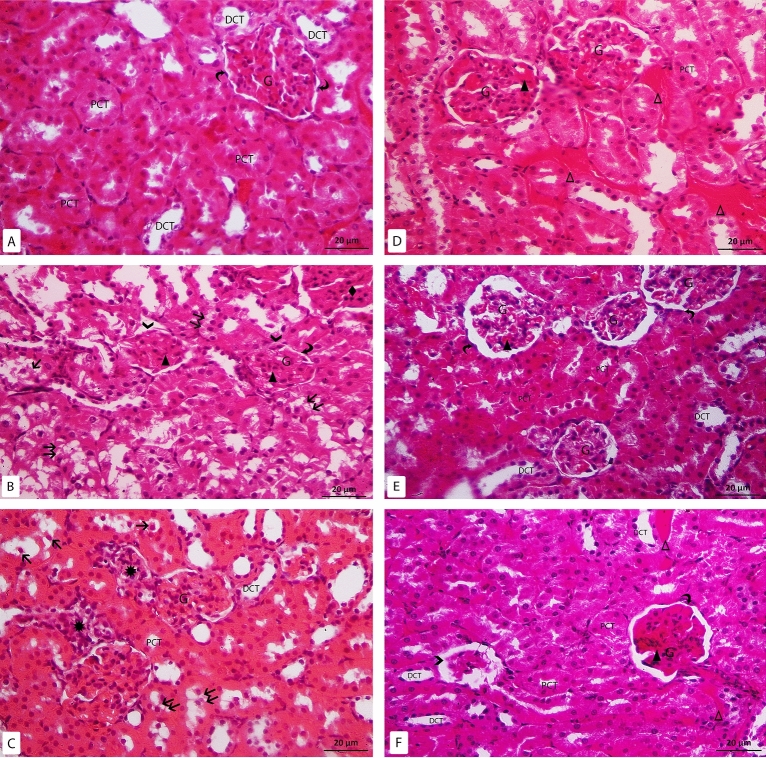
Figure 2.
H&E-stained sections X400: (A) control group: the glomerulus (G), parietal layer of Bowman’s capsule (curved arrow), proximal convoluted tubules (PCT), distal convoluted tubules (DCT). (B–D) Model-RF group: the glomerulus (G), parietal layer of bowman’s capsule (curved arrow), disrupted parietal layer of Bowman’s capsule ( >), proximal convoluted tubules (PCT), distal convoluted tubules (DCT), sloughed tubular cells (↑), vacuolated tubular cells (↑↑), mononuclear cell infiltration (*), congested glomerular (▲) and peritubular capillaries (Δ), acidophilic area with pyknotic nuclei (♦). (E,F) RF-ZA group: the glomerulus (G), parietal layer of bowman’s capsule (curved arrow), proximal convoluted tubules (PCT), distal convoluted tubules (DCT), shrunken glomerulus ( >), congested glomerular (▲) and peritubular capillaries (Δ).

