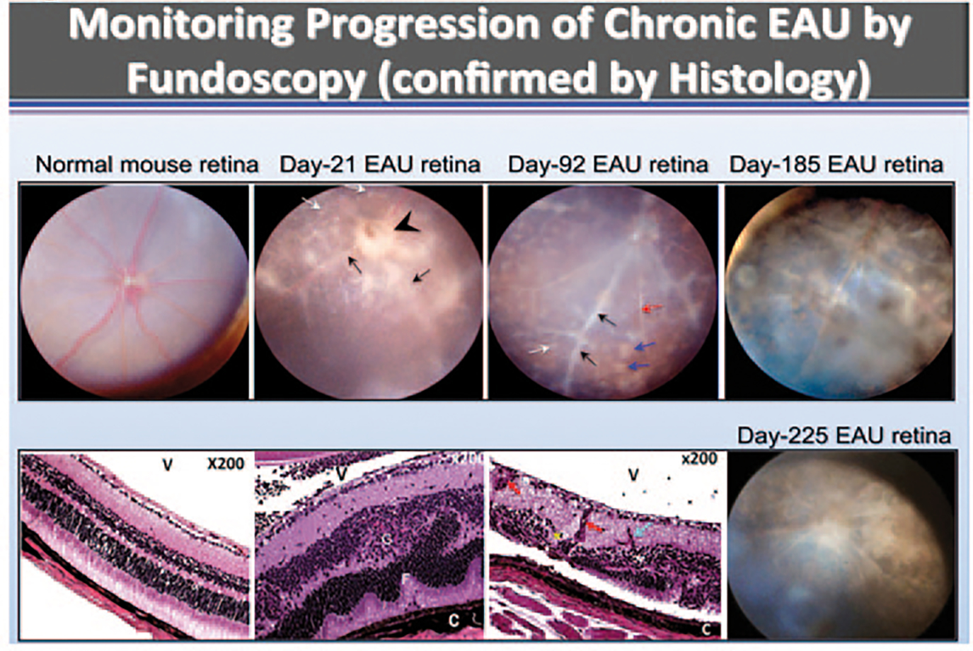
Fig. 53.1.

Chronic intraocular inflammation induces retinal degeneration and neovascularization. Fundus images were taken from normal or EAU mice over a 225 days period (top panels). Fundus images reveal blurred optic disc margins and enlarged juxtapapillary area (black arrowhead), retinal vasculitis with moderate cuffing (black arrows) and yellow-whitish retinal and choroidal infiltrates (white arrows). Histological analysis (Lower panels) reveals retinal structural damage, including evidence of atrophic retina (thinning) and sclerotic vessel (red arrow) with multiple whitish infiltrates (white arrow) and brownish chorioretinal scars (blue arrows), photoreceptor cell loss (red asterisk), retinal vasculitis (black arrows), retinal sclerotic vessel (white arrow), choroiditis (black arrowhead), and retinal degeneration (bottom panels). OpN optic nerve, V vitreous, R retina, GCL ganglion cell layer, INL inner nuclear layer, ONL outer nuclear layer, RPE retinal pigment epithelial layer, CH choroid
