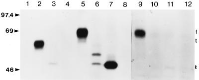
FIG. 2.
Autoradiograph of immunoprecipitated forms of membrane-anchored, secreted, and cytosolic BHV-1 gD. Preconfluent COS-7 cells transiently transfected with pSLRSV.AgD, pSLRSV.SgD, pSLRSV.CgD, or pSLRSV.Nul were grown for 48 h in the presence of 35S-labelled methionine and cysteine. Radioactively labelled gD was immunoprecipitated from medium and/or cell lysates by using an anti-gD monoclonal antibody pool (see Materials and Methods). SDS-PAGE of precipitates demonstrates the calculated molecular masses (kDa) of mutated gD and cellular localization as predicted. Immunoprecipitates of media collected from COS-7 cells transfected with the following are shown: 1, pSLRSV.AgD; 2, pSLRSV.SgD; 3, pSLRSV.CgD; and 4, pSLRSV.Nul. Immunoprecipitates of lysates of COS-7 cells transfected with the following are shown: 5, pSLRSV.AgD; 6, pSLRSV.SgD; 7, pSLRSV.CgD; and 8, pSLRSV.Nul. Immunoprecipitates of plasma membrane-associated gD are shown: 9, pSLRSV.AgD; 10, pSLRSV.SgD; 11, pSLRSV.CgD; and 12, pSLRSV.Nul. Molecular mass markers are indicated on the left. The positions of membrane-anchored (f), secreted (t), and cytosolic (c) versions of gD are indicated on the right.

