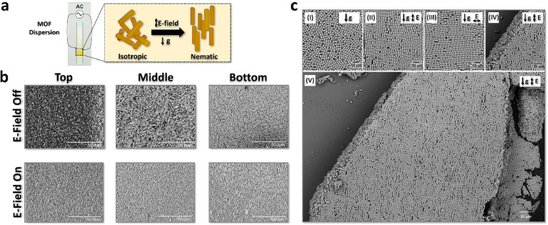
Figure 5.
a) Schematic illustration of isotropic and nematic phases of MIL-68(In) dispersion (AR = 6.8) in capillary before and after E-field-assisted sedimentation. b) Corresponding SEM images of MIL-68(In) assemblies at the top, middle, and bottom of the sediment in the absence and presence of an E-field. c) SEM images of opened capillary samples containing short MIL-68(In) (AR = 1.2) sedimented (I) without an E-field; (II) in a vertical E-field; (III) in a horizontal E-field; (IV) in a vertical E-field showing the alignment of lower particle layers; (V) in a vertical E-field showing the long-ranged order of the particles. Adapted with permission from ref (1) under a Creative Commons CC-BY license.

