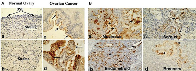
Fig. 4.
MUC13 expression in ovarian cancer was evaluated by immunohistochemistry. A, TMA. For immunohistochemical analysis, TMA slides containing ovarian carcinoma were processed. Analysis of stromal tissue and OSE from a healthy ovary revealed no detectable MUC13 expression (a, b). MUC13 (arrows) was detected in ovarian cancer samples via membrane-bound (c) and cytoplasmic (d) staining. B, Clinical tissues. In a variety of EOC samples, the expression of MUC13 was observed. Membrane-bound (a, b) and cytoplasmic (c, d) immunostaining for MUC13 in EOC samples are depicted in the four representative panels. Magnification at the outset was 200x. (Adopted with permission from ref [84])

