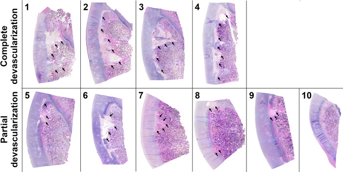
Fig 6. Representative histological images of all 10 piglets obtained 12 weeks after they underwent complete or partial devascularization of the lateral trochlear ridge of the femur.
Complete devascularization in Piglets # 1, 2, 3 and 4 led to the formation of extensive OC-manifesta lesions involving most of the lateral trochlear ridge. After partial devascularization, Piglet # 6 developed a medium sized OC-manifesta lesion involving a substantial portion of the epiphyseal cartilage; Piglets # 5, 7, 8, and 9 had healing OC-manifesta lesions nearly completely surrounded by bone; and Piglet # 10 had no apparent lesion on histology. Lesions are marked with black arrows.

