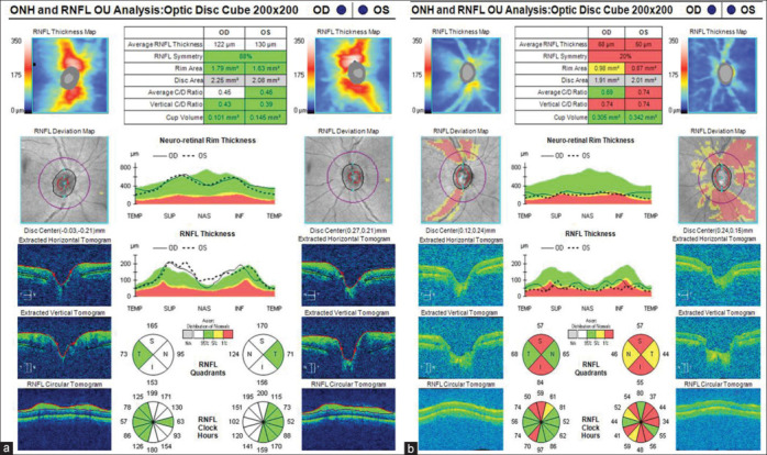
Figure 4.

Optical coherence tomography (OCT) of retinal nerve fiber layer (a) taken during first week of acute angle closure showing increased nerve fiber layer thickness due to disc edema. (b) OCT taken during last visit showing significant thinning in superior and inferior quadrants consistent with glaucomatous damage
