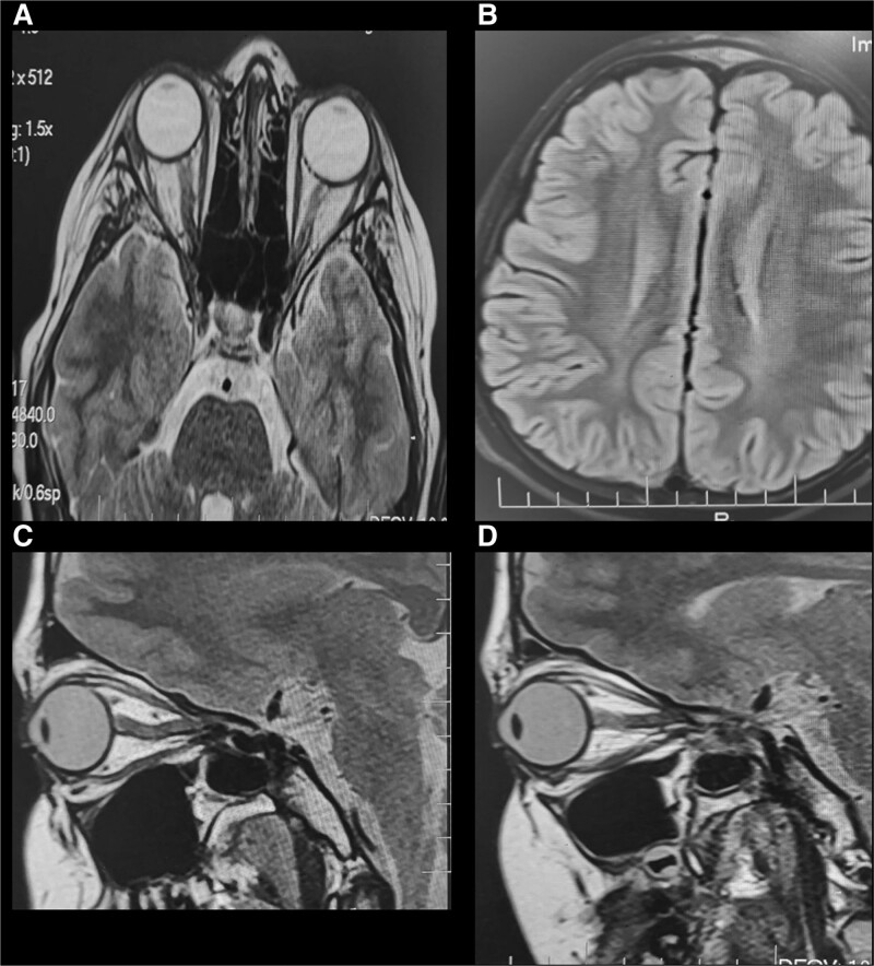
Figure 2.
(A) MRI/T2 FLAIR showed evidence of abnormal areas of peripheral enhancement, and increased optic nerve tortuosity, along the course of right optic nerve. (B) Normal brain parenchymal. No lesions (C and D) with some asymmetric right more than left (B). MRI = magnetic resonance imaging. FLAIR = fluid-attenuated inversion recovery.

