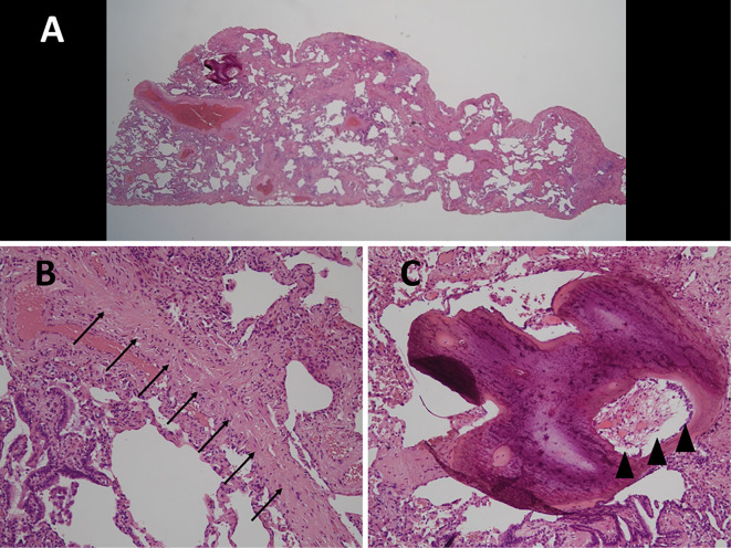
Figure 2.
Biopsy specimens obtained from the right lower lobe show relatively homogeneous thickening of the alveolar septa with fibrosis and infiltration of inflammatory cells consistent with non-specific interstitial pneumonia [A, Hematoxylin and Eosin (H&E) staining, ×1]. Findings of cicatricial organizing pneumonia such as dense fibrosis with formation of intraluminal eosinophilic scar tissue without destruction of the underlying lung architecture (B, arrow; H&E staining, ×400) and intraluminal pulmonary ossification containing bone marrow such as fat and hematopoietic cells (C, arrowhead, H&E staining, ×400) are seen.

