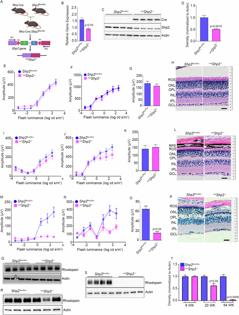
Fig. 2. Functional and morphological characterization of conditional rod-specific Shp2 KO mice.
A Rod-specific Shp2 KO mice (rodShp2−/−) were generated by breeding floxed Shp2 mice (Shp2flox/flox) with mice expressing Cre-recombinase under the control of rhodopsin promoter. Quantitative real-time PCR shows the normalized Shp2 expression to actin in Shp2flox/flox and rodShp2−/− mice (B). Data are mean ± SEM (n = 3). Retina lysates were prepared from one-month-old mice Shp2flox/flox and rodShp2−/− mice and immunoblotted with Cre, Shp2, and actin antibodies (C). Densitometric analysis of Shp2 normalized to actin (D). Data are mean ± SEM (n = 3). Functional characterization of rodShp2−/− mouse retina. Scotopic a-wave (E, I, M), scotopic b-wave (F, J, N), and photopic b-wave (G, K, O) analyses were performed on 6-week-old (E–G), 20-week-old (I–K), and 64-week-old (M–O) Shp2flox/flox and rodShp2−/− mice. Data are mean ± SEM (n = 8). The rodShp2−/− response was significantly lower than that of the Shp2flox/flox retinas (p < 0.001). Data were analyzed by two-way ANOVA and unpaired t-test. Six-week (H), 20-week (L), and 64-week-old (P) Shp2flox/flox and rodShp2−/− mouse eye sections were stained with hematoxylin and eosin staining. Retinal proteins from 6-week (Q), 20-week (R), and 64-week-old (S) Shp2flox/flox and rodShp2−/− mice were immunoblotted with rhodopsin and actin antibodies. Densitometric analysis of Shp2 normalized to actin (T). Data are mean ± SEM (n = 4 for 6 and 64 weeks; n = 3 for 20 weeks). Panels (A) was created with BioRender.com. Note: We used the same actin blot in Figs. 2R, 3D.

