
Fig. 3.
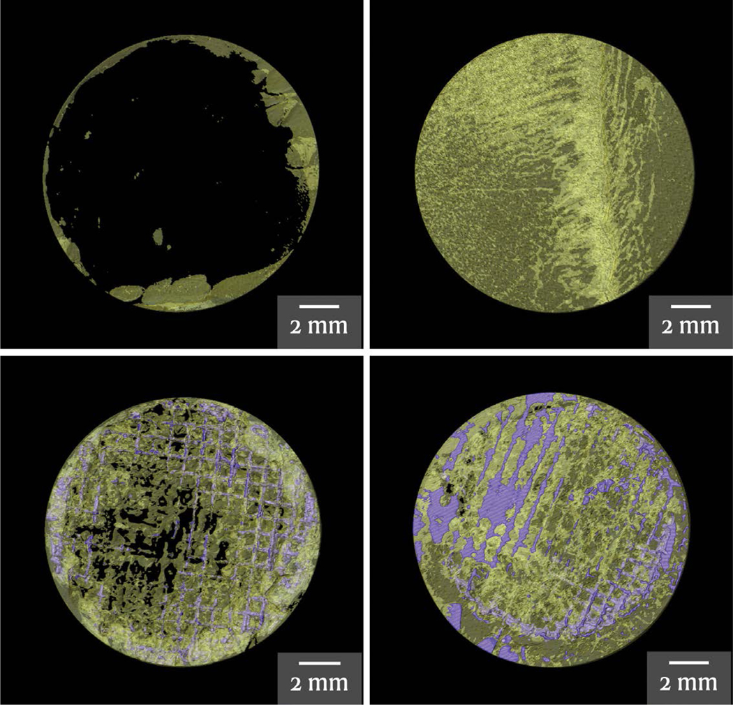
Three-dimensional reconstruction of the bone (yellow) and remaining scaffold (purple) in the defect site after 12 weeks of healing in a negative control (above, left), a sample of native bone (above, right), a defect that had been filled with a scaffold without a cap (below, left), and a defect that had been filled with a scaffold with a cap (below, right).