
Extended Data Figure 7: Comparative protein structure modelling of an integrase family sequence supports annotation as a tyrosine recombinase.
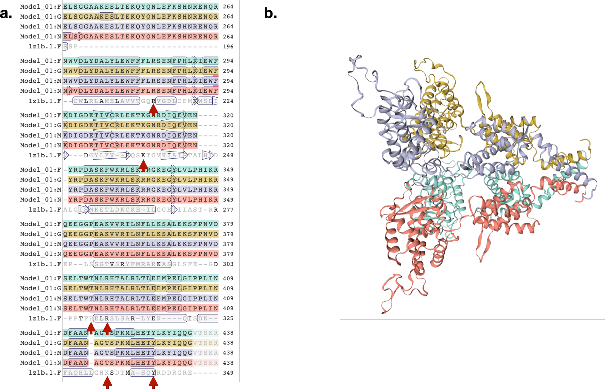
(a) Target/template alignment between the Prochlorococcus PAC1 sequence (indicated as Model _01), and the template sequence 1Z1B, the phage lambda integrase. Red arrows point to active site residues Arg 212, Lys 235, His 308, Arg 311, His 333, and Tyr 342. Boxed amino acid regions represent secondary structure. (b) Homology model of Prochlorococcus PAC1 sequence based on template 1Z1B. Colors indicate individual monomers of the homo-tetramer template protein structure in both a and b.