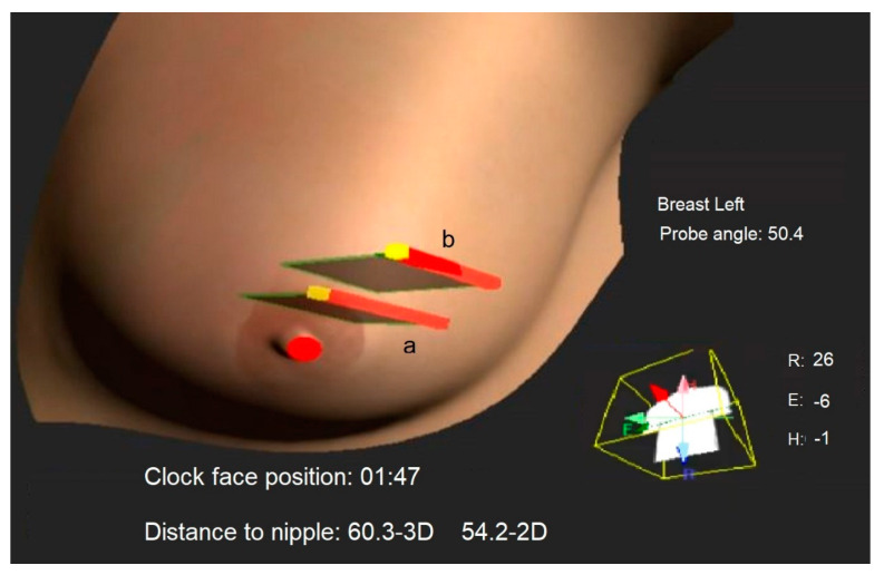
Figure 1.
The 3D ultrasound frame position and orientation from a previous exam, labeled as ‘a’, is displayed as a gray rectangle. This is shown concurrently with the real-time ultrasound frame during the current exam, labeled as ‘b’. This approach assists consecutive operators in reproducing the 3D mass location by aligning frame ‘b’ with frame ‘a’. The probe head is represented by a red line, the orientation marker by a yellow dot and the nipple point by a red dot. Additionally, metrics about the current probe position relative to the nipple and body, as well as the body’s rotation on the exam table, are displayed in real-time for operator guidance.

