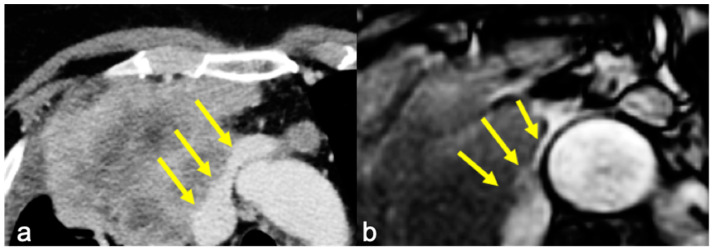
Figure 2.
(a) Axial contrast-enhanced CT showing irregular margins at the left innominate vein and superior vena cava confluence and (b) cine-MRI of the same patient with no “India Ink” artifact between the mass and the vessels, suspicious for infiltration (yellow arrows). Infiltration of the left brachiocephalic vein was confirmed at surgery.

