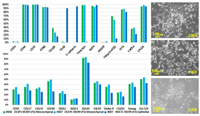
Figure 2.
Exosome characterization of cell lines. (right) These are phase-contrast images of #006 and #007 human ovarian carcinoma ascites (upper and middle) and #007 human ovarian carcinoma tissue (lower)-derived cells (P2). The adherent culture conditions were M199 + 10% FBS + 20 ng/mL of EGF + 0.4 μg/mL of hydrocortisone. (left) These are surface expression markers of human ovarian carcinoma ascites and tissue-derived cells with spindle-like mesenchymal-like (MSC-) (right upper and middle) ovarian carcinoma stromal progenitor cells (OCSPCs) and roundish epithelial-like (epi-) (right lower) ovarian-carcinoma-tissue-derived cells from 2 advanced ovarian cancer patients. (left) High expressions of vimentin in MSC-OCSPCs and CK18 and E-cadherin in epi-OCSPCs were noted. High expression of CD44, CD73, CD90, FLT4, CA125, and SSEA4 was noted in both cells.

