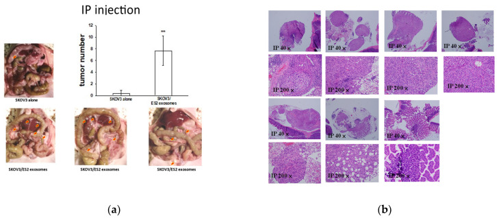
Figure 9.
EOC-derived EXs accelerated cancerous peritoneal dissemination. (a) Representative pictures of 6/7 mice IP injected with 1 × 10⁶ SKOV3 cells with ES2 exosomes showing disseminated tumors (red arrows) in the peritoneal cavity compared to the 1/3 mice injected with 1 × 10⁶ SKOV3 cells with PBS (p = 0.097, as determined using Student’s t-test). The average disseminated tumor numbers in the peritoneal cavity were significantly greater in mice receiving SKOV3 cells with ES2-exosomes than in those administered SKOV3 cells with PBS (** p < 0.01, as determined using Student’s t-test). (b) Representative histologic pictures of disseminated peritoneal tumors are shown at microscopic scales of 40× and 200×.

