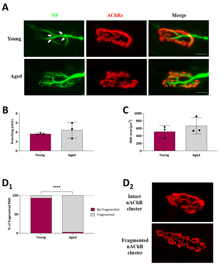
Figure 2.
NMJ morphological analysis of young and ageing EDL muscles. (A), Immunofluorescence microscope images of representative NMJs from young and ageing EDL muscles, doubly labelled to identify the presynaptic nerve terminal (neurofilament, green), and the postsynaptic end plate (nAChRs; α-bungarotoxin, red). White arrows indicate the branching points. (B–D1), Histograms showing the quantification of morphological analysis. (B) Quantification of the number of branching points, (C) postsynaptic NMJ area, and (D1) NMJ fragmentation. Fragmented nAChR clusters are defined as five or more separated islands of nAChR clusters to form the endplate (previously described in [28]). Note that the percentages of fragmented NMJ significantly increase in ageing muscles. (D2) Representative images of intact and fragmented nAChR clusters showing how most young and ageing NMJs look, respectively. Scale bar: 25 µm. Data are represented as means ± SD. Statistical significance was determined using the Mann–Whitney test for graphs (B,C), and the graph (D1) was determined by 2-way ANOVA (**** p value < 0.0001). A minimum of 30 NMJs per rat from at least three separate rats of each group were quantified.

