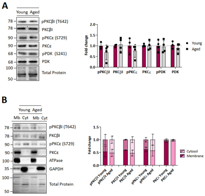
Figure 5.
Protein kinase C and PDK levels of young and ageing EDL muscles. (A), Western blot analysis of different protein kinases and their phosphorylated forms. Quantification analysis shows that cPKCβI, nPKCε, and PDK1 protein levels (and their respective phosphorylated active forms) do not change in ageing muscles. (B), Distribution of the protein kinases between the membrane and cytosol fractions. PKCs are equally distributed in young and ageing muscles. Data are represented as means ± SD. Statistical significance was determined using the Mann–Whitney test.

