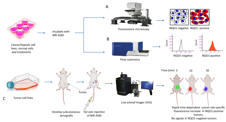
Figure 11.
(A) Diagrammatic summary of techniques for NQO1 imaging using NIR-ASM and resulting fluorescence in NQO1-positive cancer cells and lack of it in NQO1-negative (normal) cells after incubation of 10 µM NIR-ASM for 1 h is shown. (B) Flow cytometry assays to determine NIR-ASM activation by NQO1-positive and -negative cell lines (C) Application of live in vivo fluorescence imaging of NQO1-positive tumors developed in nude mice.

