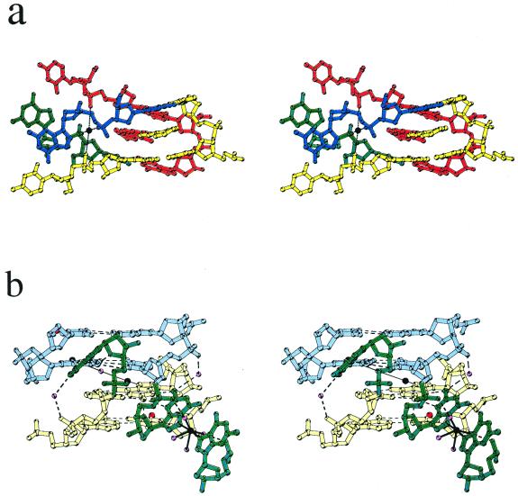
Figure 4.
(a) Stereo view of the double intercalation site of the d(CGTACG) complex. DNA and corresponding 9-aminobutyl-DACA molecules from different duplexes are represented in different colours. Nucleotides C7 and G8 from four different duplexes are shown, nucleotides C5 and G6 for two duplexes only (yellow and red) are drawn. The sodium ion is drawn in black. Black dashed lines indicate sodium coordination bonds to the guanine G8 phosphate oxygens. (b) Stereo view of the unpaired end of the d(CG5BrUACG) showing the Co(II) ion coordinated to the N7 atoms of two guanine G12s. The G12 nucleotides are coloured green, Co(II) ions black, bromine atoms red and water oxygen atoms pink. Nucleotide pairs A10/U3 and C11/G2 from the two duplexes are shown in different colours (blue and yellow) to distinguish the different duplexes. Dashed lines indicate hydrogen bonds and solid black lines indicate Co(II) coordination bonds.

