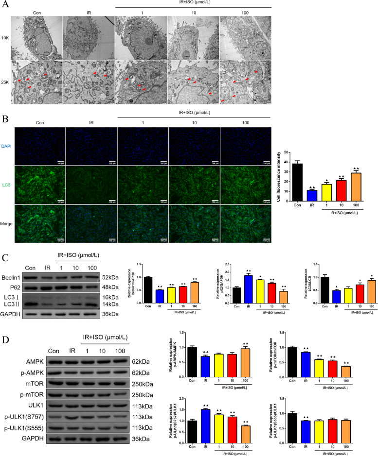
Fig. 4.
ISO treatment caused autophagy induction by AMPK/mTOR/ULK1 pathway in IR H9c2 cells. A The detection of H9c2 cell ultra-structure was completed by TEM (magnification 10.0 k × , scale bar: 2 μm; magnification 25.0 k × , scale bar: 500 nm), red arrows showed autophagosome, n = 6; B The immunofluorescence assay was conducted to measure LC3 fluorescence expression in H9c2 cells (magnification 200 × , scale bar: 100 μm), n = 6; C Western blot was employed to measure Beclin1, P62 and LC3II/LC3I protein expression in H9c2 cells, n = 3; D The p-AMPK/AMPK, p-mTOR/mTOR, and p-ULK1(S757)/ULK1, and p-ULK1(S555)/ULK1 protein expression of H9c2 cells was tested by western blot assay, n = 3; ▲P < 0.05 and ▲▲P < 0.01 vs. Con group, ★P < 0.05 and ★★P < 0.01 vs. IR group. Note: Con: Control; IR: Ischemia reperfusion; ISO: Isoliquiritigenin; TEM: Transmission electron microscopy

