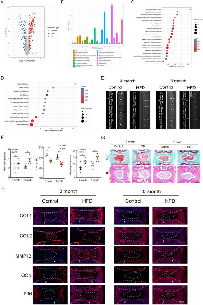
Fig. 3.
HFD (High Fat Diet) induces cartilage endplate cell senescence and calcification in rat intervertebral discs. A Differentially expressed protein volcano maps from quantitative proteomics. B COG /KOG functional classification. C, D GO enrichment analysis of differentially expressed proteins (biological processes and cellular components) derived from quantitative proteomics analysis. E Representative micro-CT and MRI images of caudal vertebrae from rats fed with HFD for 3 and 6 months (n = 5). White and yellow arrows indicate the target disc. F Statistical plot of Pfirrmann grade, DHI and histological scores (n = 5). G Representative images of HE, SO staining of the rat coccygeal spine IVD (n = 5). Scale bar = 500 μm. H Immunofluorescence representative images of MMP13, COL2, P16, OCN and COL1 (n = 5). Scale bar = 500 μm. White arrow: cartilage endplate. Compared to control group, *p < 0.05, **p < 0.01, ***p < 0.001

