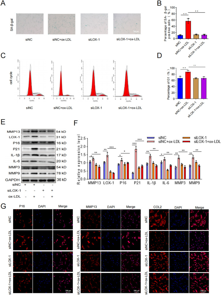
Fig. 6.
The induction of senescence in cartilage endplate cells by ox-LDL was mediated through LOX-1. A, B Results of SA-β-Gal staining and quantitative analysis (Scale bar = 200 μm). C, D Cell cycle detection and analysis results. E, F Western blotting and quantitative analysis of LOX-1, MMP13, p16INK4α, P21, IL-1β, IL-6, MMP3 and MMP9. G-I COL2, MMP13, P16 representative immunofluorescence staining results. Compared to siNC + ox-LDL group, *p < 0.05, **p < 0.01, ***p < 0.001

