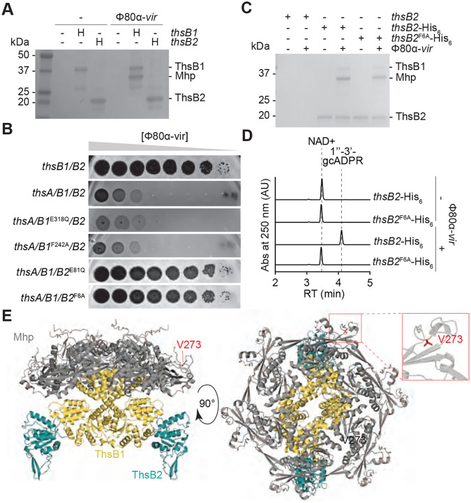
Figure 4. ThsB1 interacts with Mhp to recruit ThsB2 and stimulate its cyclase activity.
(A) Coomassie Blue-stained SDS-PAGE of proteins isolated from staphylococci expressing hexahystidyl- (H) tagged versions of ThsB1 or ThsB2, uninfected or infected with Φ80α-vir, after cobalt resin affinity chromatography. Protein molecular weight (kDa) markers are shown. (B) Tenfold serial dilutions of Φ80α-vir on lawns of S. aureus RN4220 harboring plasmids carrying either an incomplete (thsB1/B2) or full (thsA/B1/B2) Thoeris operon carrying wild-type or mutant versions of thsB1 or thsB2. (C) Coomassie Blue-stained SDS-PAGE of proteins isolated from staphylococci expressing ThsB2, ThsB2-His6 or ThsB2F6A-His6, uninfected or infected with Φ80α-vir, after cobalt resin affinity chromatography. Protein molecular weight (kDa) markers are shown. (D) HPLC analysis of the products resulting from the incubation of the proteins purified from staphylococci expressing ThsB2-His6 or ThsB2F6A-His6, uninfected or infected with Φ80α-vir, with NAD+, using a cobalt resin. Retention times (RT) of reactants and products are marked by dotted lines. (E) AlphaFold3 structure of a complex formed by Φ80α Mhp (hexamer; grey) ThsB1 (two copies; yellow) and ThsB2 (two copies; teal). Two angles of the structure (90° rotation), as well as the position of residue V273 (red) are shown.

