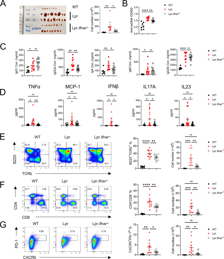
Figure 3: IFNAR1 deletion reduces systemic inflammation in Lpr mice.
(A) Representative image of peripheral lymph nodes (left) and cellularity (right) from 6 months B6, Lpr, and Lpr.Ifna1r−/− mice. (B) Tiers of anti-ssDNA antibody level in mice serum measured by ELISA. Serum immunoglobulin (Ig) isotype concentration (C) and inflammatory cytokine concentration (D) in 6-month-old mice. (E) Expression of TCRb and B220 in pLN derived lymphocytes. Left, frequency of TCRb+B220+ population; right absolute cell number of TCRb+B220+ cells in pLN. (F) Expression of CD4 and CD8 in pLN derived lymphocytes. Left, frequency of CD4−CD8− population; right absolute cell number of CD4−CD8− cells in pLN. (G) Expression of CXCR5 and PD-1 in pLN derived lymphocytes. Left, frequency of CXCR5+PD-1hi Tfh cells; right, absolute cell number of CXCR5+PD-1hi Tfh cells in pLN. ns, not significant; *p < 0.05, **p < 0.01, ***p < 0.001, ****p < 0.0001. p-Values were calculated with one-way ANOVA with post hoc Tukey test. Error bars represent SEM.

