Figure 2.
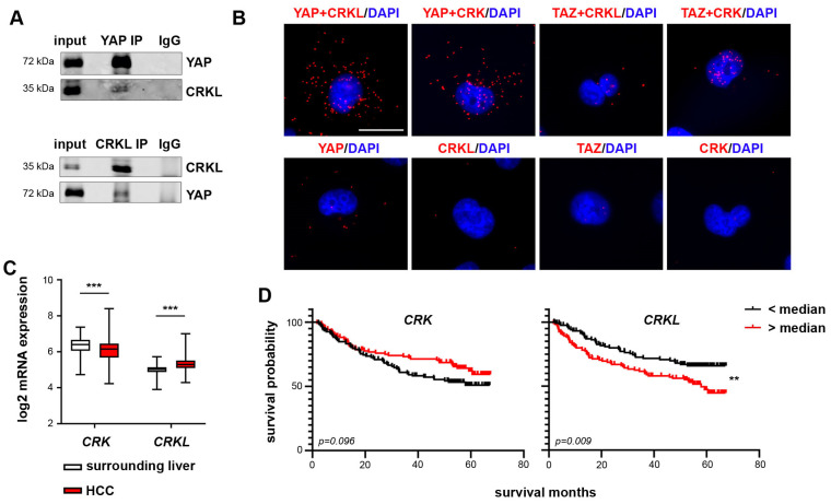
CRK and CRKL are new YAP interaction partners. (A) Precipitation of either endogenous YAP (upper panel) or CRKL (lower panel) and co-precipitated CRKL or YAP in HLF cells, respectively, confirming the interaction between both proteins. IgG served as negative control. (B) In situ PLA confirmed the predicted interactions between YAP and CRK/CRKL and TAZ and CRK in HLF cells. For TAZ and CRKL, no clear interaction was detected. Negative controls used only one primary antibody for PLA. Scale bar: 20 µm. (C) The transcriptome data of HCC tissue and the corresponding surrounding liver tissue (N = 242) [29] were analyzed for the mRNA expression of CRK and CRKL. While the CRK expression was decreased, CRKL expression was increased in HCC tissue. Statistical test: Mann–Whitney-U. (D) Patients were stratified into high- and low-expression groups using the median as a cutoff. Kaplan–Meier plots show an increased survival probability for patients with high CRK expression and a significantly decreased probability for patients with high CRKL expression. Statistical test: log-rank test. p ≤ 0.01 **, p ≤ 0.001 ***.

