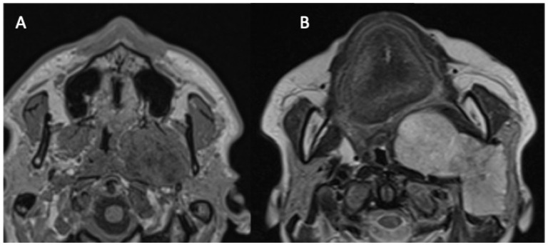
Figure 1.
Two forms of the parapharyngeal space involvement of parotid tumors. Magnetic resonance images: axial T1WI images at the level of the hard palate. (A) Non-contrast image presenting an isolated PPS tumor developed from the medial protuberance of the parotid gland deep lobe. (B) Contrast-enhanced image showing a deep lobe tumor with the extensive involvement of the PPS.

