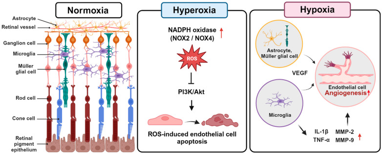
Figure 1.
Cellular mechanisms involved in the development of retinopathy of prematurity (ROP). During hyperoxic conditions, phase 1, nitro-oxidative stress induces apoptosis in retinal capillary endothelial cells by disrupting the PI3K/Akt signaling pathway. During hypoxic conditions, phase 2, astrocytes and Müller glial cells secrete VEGF, while microglia secrete IL-1β, TNF-α, and metalloproteinases, promoting neovascularization by endothelial cells.

